Describe the Structure of the Heart
Cardiac dynamics are traditionally linked to a left ventricle right ventricle and septum morphology a topography that differs from the hearts five-century-old anatomic description of containing a helix and circumferential wrap architectural configuration. The heart is enclosed in a tough two-layered sac the pericardium comprising inner visceral pericardium attached to the heart and the outer parietal pericardium.

Pin On Studying
The diagram of heart is beneficial for Class 10 and 12 and is frequently.
. During the time that the SA Node has the electricity the Atria in the heart contract. The ventricles are the chambers that pump blood and atrium are the chambers that receive blood. The human heart is the most crucial organ of the human body.
The three layers of the heart as discussed above are the epicardium. Structure of the Heart. And the lower two chambers are called the right and left ventricle.
The electrical impulse travels from the sinus node to. The upper two chambers are called right and left atrium. The heart measures 12 x 85 x 6 cm and weighs 310 g males and 255 g females Relations.
It is located in the middle cavity of the chest between the lungs. It is positioned posteriorly to the body of the sternum with one-third situated on the right and two-thirds on the left of the midline. The heart has a somewhat conical form and is enclosed by the pericardium.
The shape of the heart is similar to a pinecone rather broad at the superior surface and. The heart consists of four chambers right and left atria above right and left ventricles. The heart is made up of four chambers.
The human heart is divided into four chambers. The bottom is shaped like a v. These layers work together to form the major structure of the heart and allow it to function after endless hours of pumping.
The tricuspid pulmonary mitral and aortic valves. A small body of specialized muscle tissue in the wall of the right atrium of the heart that acts as a pacemaker by producing a contractile signal at regular intervals. The two layers have a potential space or cavity in between them the pericardial cavity which consists of about 50 ml of pericardial fluid.
The heart wall is divided into three layers. Recall that the hearts contraction cycle follows a dual pattern of circulationthe pulmonary lungsand systemic body circuitsbecause of the pairs of chambers that pump blood into the circulation. Each side of the heart consists of an atrium and a ventricle which are two connected chambers.
It is the cardiac muscle that enables the heart to contract and allows for the synchronization of the heartbeat. The coronary vessels that serve the heart pulmonary heart and lungs and systemic systems of the body. The electrical stimulus travels down through the conduction pathways and causes the hearts ventricles to contract and pump out blood.
This is a fibrous covering that wraps around the heart and holds it in place. Epicardium myocardium and endocardium. Chambers and Circulation through the Heart.
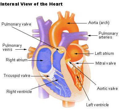
Internal Structure of the Heart. The mitral valve is an atrioventricular valve. Among which both right atrium and ventricle make up the right heart and the left atrium and ventricle make up the left heart The structure of the heart also houses the biggest artery in the body the aorta.
This valve prevents backflow of the blood into the atrium above when the ventricle contracts. The most common heart attack symptoms or warning signs are chest pain breathlessness nausea sweating etc. Two atria on the top and two ventricles on the bottom.
It is also made up of four valves. Shape and Size of the Heart. In order to develop a more precise understanding of cardiac function it is first necessary to explore the internal anatomical structures in more detail.
Both the aortic and the pulmonary valves are semilunar valves. Heart is the organ in the human body that acts as a double pump. In most people the heart is located on the left side of the chest beneath the breastbone.
It pumps blood from the heart to different parts of the body and back to the heart. The heart consists of four chambers. The left atrium receives the oxygenated blood from the lungs via the pulmonary vein.
The heart is composed of smooth muscle. The mitral valve separates the left ventricle from the left atrium. The right atrium links to the right ventricle by the tricuspid valve.
The 2 upper chambers of the heart atria are stimulated first and contract for a short period of time before the 2 lower chambers of the heart ventricles. The human heart is located within the thoracic cavity medially between the lungs in the space. The outer protective layer of the heart.
Parts of the human heart. Two upper chambers known as the left atrium and right atrium and two lower chambers called the left and right ventricles. Looking at the Valentines Day heart the two rounded humps at the top are rounded like the top of a lower-case a.
Coronary circulation intrinsic to the heart takes blood directly from the main artery aorta coming from the heart. The functions of each part are as follows. The heart wall is composed of connective tissue endothelium and cardiac muscle.
Heart Anatomy Location of the Heart. The heart is a muscular organ that pumps blood throughout the body. Heart structure and function are closely related as described below.
Known as the P Wave on an EKG. Sign up for our weekly newsletter. The heart is a complex muscle that pumps blood through the three divisions of the circulatory system.
STRUCTURE OF HUMAN HEART. It sends electricity currents to the AV Node in the heart. It has four chambers which contract in a specific order allowing the human heart to pump blood.
Blood travels through the bicuspid valve to the left atrium. The atria plural of atrium are where the blood collects when it enters the heart. Although it is barely the size of a human fist the heart is a powerful muscle inside the chest with a cone shape and a pointed end facing the left.

How The Human Heart Evolved Four Chambers Human Heart Diagram Heart Anatomy Heart Diagram
Seer Training Structure Of The Heart

Http Www Aviva Co Uk Library Images Med Encyclopedia Cfhg350strhea 002 Gif Heart Structure Human Anatomy And Physiology Anatomy And Physiology

Heart And Great Vessels Anterior View The Heart Is Enclosed In The Pericardial Sac The Innermost Layer Of W Heart Structure Cardiovascular System Emt Study


Comments
Post a Comment